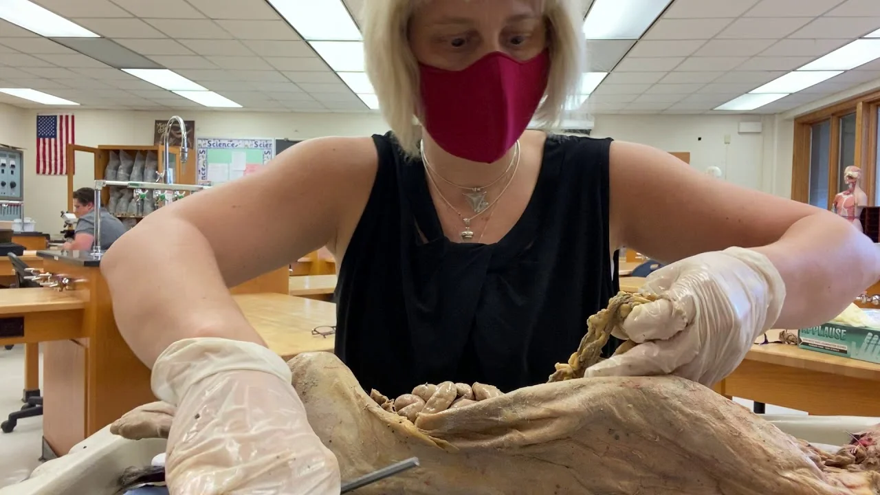
Dr. Molescu, assistant professor at Daytona State College, continues the cat dissection by removing and examining the major organs of the thoracic and abdominal cavities.
Thoracic Cavity: Lungs, Trachea, and Diaphragm ⏱ 0:33
•The thoracic cavity contains right and left pleural cavities holding the lungs.•The right lung and left lung are dissected out using a ten blade and scalpel.•After lung removal, the mediastinum contains the inferior vena cava and aorta (dyed blue and red respectively).•The trachea is isolated and shown with tracheal rings, which feel cartilaginous.•The diaphragm is a dome-shaped structure located over the liver.•The inferior vena cava pierces through the diaphragm.Abdominal Organs: Liver, Gallbladder, Pancreas, and Stomach ⏱ 11:34
•The liver is removed along with the gallbladder underneath; the hepatic artery is identified.•The inferior vena cava runs through the liver; a probe is placed through it.•The gallbladder is a small structure located under the liver.•The greater omentum and lesser omentum are dissected; the pancreas is found retroperitoneal behind the stomach.•The stomach is dissected with the first part of the small intestine (duodenum/duodenum).•The stomach is J-shaped; the duodenum is the first part of the small intestine, followed by jejunum and ilium.Small and Large Intestine, Esophagus ⏱ 14:43
•The esophagus is dissected; it is a very strong muscular tube.•A cross cut of the esophagus is shown.•The jejunum and ilium (last part of small intestine) are removed.•The cecum and large intestine are identified; the large intestine has a larger texture than the small intestine.•Poop is visible in the large intestine; it is not squeezed.Urinary System: Kidney, Ureter, Bladder, Urethra ⏱ 17:12
•The kidney has a lot of fat around it for protection; the fat is dissected away.•The ureter is identified coming out of the kidney and going to the bladder.•The bladder and urethra are dissected; the cat is female.•The renal artery and renal vein are shown coming off the aorta and vena cava.•The kidney is removed with the renal artery and renal vein attached at the hilum.•The thick fibrous renal capsule is peeled off, revealing the kidney.•A sagittal cut of the kidney is performed; renal pyramids (darker in color) and renal columns (between pyramids) are visible.•The renal artery branches into segmental, interlobar, arcuate, and corticoradiate arteries feeding into each pyramid.Key Takeaways
•The thoracic cavity contains the lungs, trachea, and diaphragm; the inferior vena cava pierces the diaphragm.•The pancreas is retroperitoneal behind the stomach; the gallbladder is located under the liver.•The esophagus is a strong muscular tube; the large intestine has a larger texture than the small intestine.•The kidney is protected by a thick fat layer; the renal capsule can be peeled off like a fascial plane.•A sagittal cut of the kidney reveals renal pyramids, columns, and the branching of the renal artery (segmental, interlobar, arcuate, corticoradiate).Conclusion
This instructional video covered the dissection and examination of major thoracic and abdominal organs, providing a hands-on review of anatomical structures.